Capsule Stain
Materials:
- microscope slide
- nigrosin stain
- safranin or crystal violet
- DI water
- inoculating loop
- Bunsen burner
Procedure:
First, we had to place a small drop of nigrosin stain onto the slide and inoculate our bacterial samples into the drop. Once they had been thoroughly mixed together, a second slide's short edge was placed at a 45 degree angle in the bacteria-nigrosin drop. The second slide was then drawn across the length of the first slide, thereby, adequately spreading out the drop. We allowed this to dry.

Once the spread smear dried, we covered it with crystal violet for approximately thirty seconds then rinsed it with DI water to remove any excess stain. The slide was then blotted with bibulous paper.

Results:
The environmental sample (above) definitely had capsules. We were able to observe using the microscope the capsules surrounding the bacteria. Our unknown (below), on the other hand, was un-encapsulated.

Endospore Staining
Materials:
- slide with fixed smear of bacteria
- staining reagents - safranin and malachite green
- steam from DI boiling water over hotplate
- staining rack
- filter paper
- DI water
- bibulous paper
- forceps
Procedure:
 We began by placing the slide (we did both our unknown and environmental) on a rack over the boiling water. A piece of filter paper was placed on the slide and saturated with malachite green. We let the specimen stain for approximately 5 minutes, adding more malachite green as it evaporated to prevent drying. We removed the slide from the heat, and placed the filter paper in the biohazard bag. The slide then cooled.
After the slide cooled, we covered the smear with safranin for 60 - 90 seconds. Afterwards, we rinsed the slide with DI water to remove excess safranin and blotted the slide with bibulous paper.
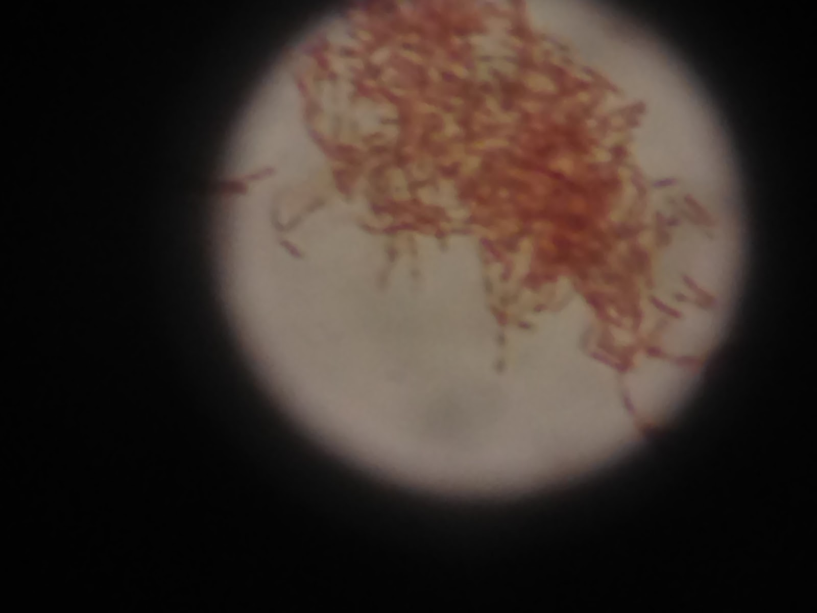
Results/application:
Our unknown endospore stain revealed that our bacteria had endospores. Our environmental picture came out blurry, but from what we can tell, it did not have endospores in it. Knowing whether a bacteria has endospores or capsules will determine what method to take to kill the bacteria since capsules and endospores can resist harsh conditions.
 |
| Unknown sample - endospore stain |
 |
| Environmental - endospore stain |
No comments:
Post a Comment